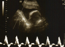
Vasa Praevia Screening
Screening for Vasa Praevia at The Brayford Studio Lincolnshire Vasa Praevia Screening

Vasa Praevia is a rare condition (1 in 3000) in which fetal blood vessels traverse the lower uterine sigment in advance of the presenting part. The vessels are not supported with the placenta or the umbilical cord. Vasa praevia presents with painless vaginal bleeding at the time of either spontaneous or at artificial rupture of membranes, ARM. Fetal shock or death may develop rapidly. Incidence of fetal death in unrecognized cases prior to onset of labour may be as high as 100%.
RISK Factors for vasa praevia:
1 - Bi-lobed and succenturate lobe placentas
2 - Low lying placenta
3 - Multiple pregnancies
4 - IVF Pregnancies
5 - Marginal and velamentous insertion of the umbilical cord
6 - History of uterine surgery or D&C
7 - Painless vaginal bleeding
Screening using transvaginal ultrasound scanning at 12, 20,28 and 34 weeks of pregnancy in high risk cases may be a useful tool to avoid uneccessary fetal morbidity and mortality in labour.
Difficulties in diagnosis of vasa praevia during ultrasound doppler Scanning may be due to:
1 - Amniotic band
2 - Chorioamniotic membrane separation
3 - Normal cord loop
4 - Marginal placental vascular sinus
Scanning may be also difficult in cases of
1 - Maternal size
2 - Maternal bladder status
3 - Orientation of the vasa praevia vessels as they cross in front of the presenting part, However Colour Doppler will differentiate all these conditions.
The Vasa Praevia appearance on the scan may be one of the following:
1 - Insertion of the umbilical cord is not into the placenta but into the attached membranes across the internal cervical os, i.e velamentous insertion OR
2 - Colour doppler may detect the intramembraneous vessels at the cervical os more readily OR
3 - Colour doppler and endo vaginal scanning can confirm the presence of aberrant vessels over the internal cervical os
Repeat examinations as recommended will avoid Flash artefact found during scanning. In all ultrasound scan examination the normal central umbilical cord placental insertion should noted and documented
The International Vasa Praevia Foundation (IVPF ) believes that fetal mortality due to vasa praevia is an avoidable tragedy. Vasa Praevia may be prevented with early diagnosis and treated with elective caesarean section performed at 35 weeks. Specific ultrasound (Colour Doppler) screening is the key for diagnosing vasa pravia
Specific ultrasound, Transvaginal Colour Doppler Screening is therefore the key for diagnosing vasa praevia from 12 weeks of pregnancy onwards. It is the state of the art technology used at the Brayford Studio Lincolnshire.
| Package | Price |
| Vasa Praevia Screening | £139.99 | 2D Placenta Praevia (Low Lying Placenta) | £125.99 |
Find out more about the following scans:


